The sacroiliac joint is small, deep, and hidden behind bony landmarks that shift with the pelvis. Whoever fixates that joint does not see the target: they infer the target from the image.
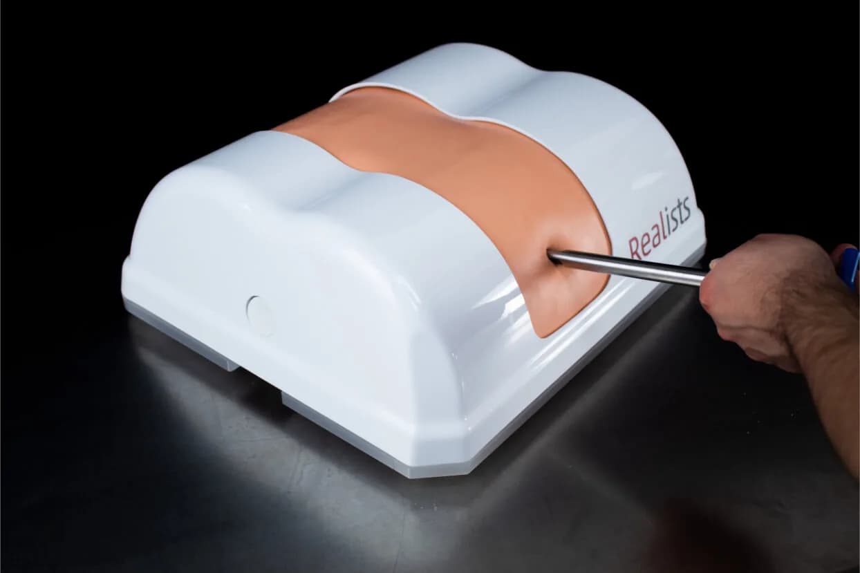
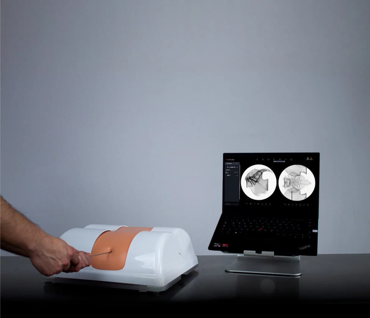
Basic SI reproduces the sacrum and the ilium from S1 to S3, with cancellous and cortical bone at distinct densities and with the roughness the drill meets. The access is percutaneous, lateral or posterior, and the image can come from X-ray, from external navigation or from RealImaging.
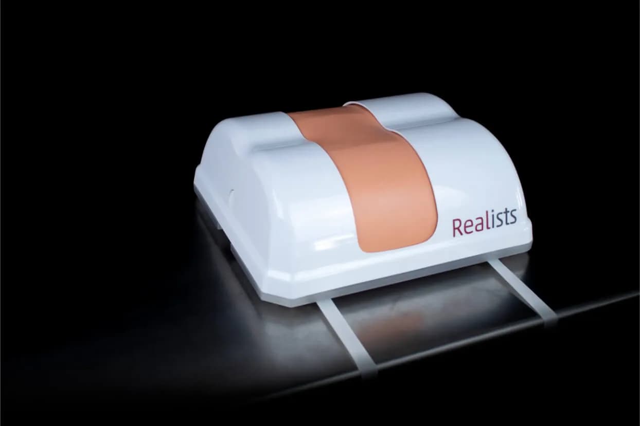
What changes between sessions is the anatomical cartridge, with disposable skin and muscle. The base and the cover are reusable, and the assembly straps to the table so it does not move during the procedure.