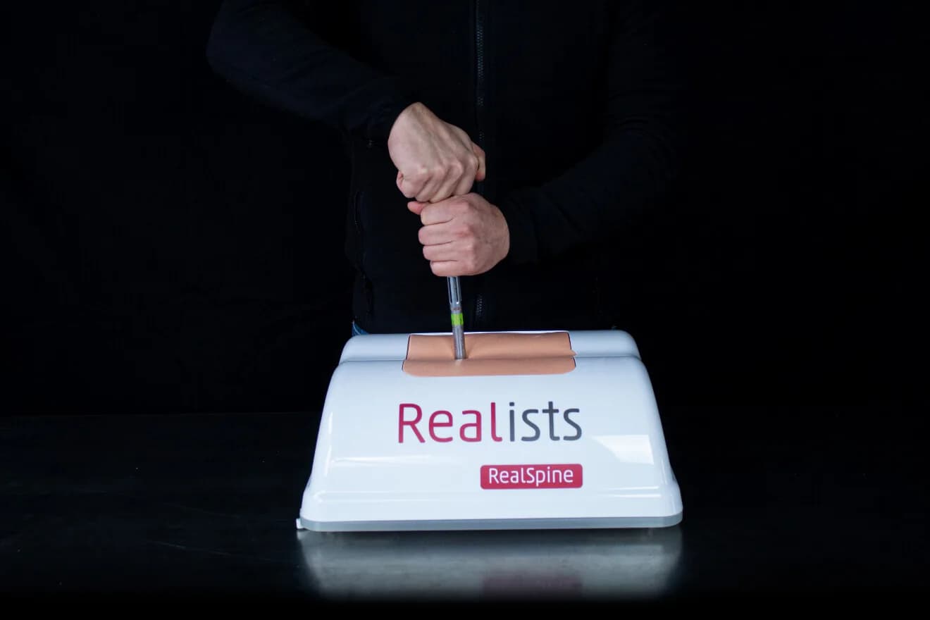
The pedicle screw is placed almost blind. The pedicle leaves a few millimetres of margin on each side, and confirmation of the trajectory comes from the image, not from the eye.
Basic Spondy X exists for that gesture. The model is radiolucent, so the X-ray shows the instrument and the screw, not the simulator. The pathology comes ready: spondylolisthesis at L4-L5, with fixation in the pedicles of L3, L4, L5 and S1.
The access is posterior, open or percutaneous, and the bone and tissues keep the behaviour of the rest of the RealSpine line. The image can come from X-ray, from external navigation, from robotics, or from RealImaging when there is no C-arm in the room.