HeartWorks teaches cardiac anatomy and echocardiography side by side, but it lives in the simulation lab: the manikin, the two probes and the station. Anyone who wants to revise an imaging plane the night before, explain an anatomical relationship to a colleague in the corridor, or show a patient what the scan found, does not carry the station along.
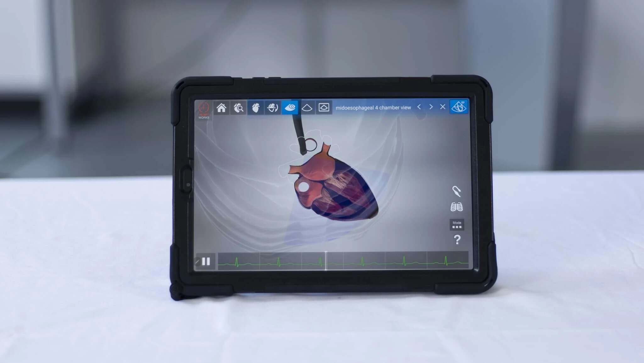
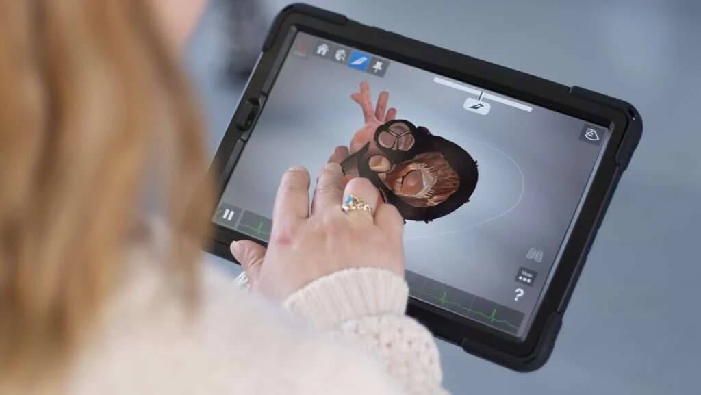
HeartWorks AR is the same three-dimensional heart, on a tablet. The model appears on screen or positioned in the room in front of whoever holds the device, to be turned in the hand and looked into. It can be zoomed, sliced along any plane, and lit up one structure at a time.
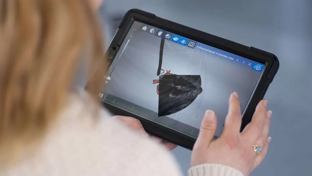
Echocardiography is still part of it. These are the reference planes for both routes, 28 transoesophageal and 20 transthoracic, with a transparent rib cage showing where the probe sits for each one.
The manufacturer designed the AR version for three quite different uses: self-directed study, tutor-led teaching, and presenting to a whole room.