

HeartWorks
Cardiac anatomy and echocardiography teaching on a three-dimensional heart of 135 structures, scanned with transthoracic and transoesophageal probes.
PresentOverview
Echocardiography is taught in two halves that rarely meet. On one side the anatomy of the heart, studied in atlases and specimens. On the other the imaging plane, learned on screen, in black and white, with the patient in front of you. A beginner has to bridge the two in their head, and that is where learning stalls: you cannot tell what you are seeing because you cannot tell where the beam went.
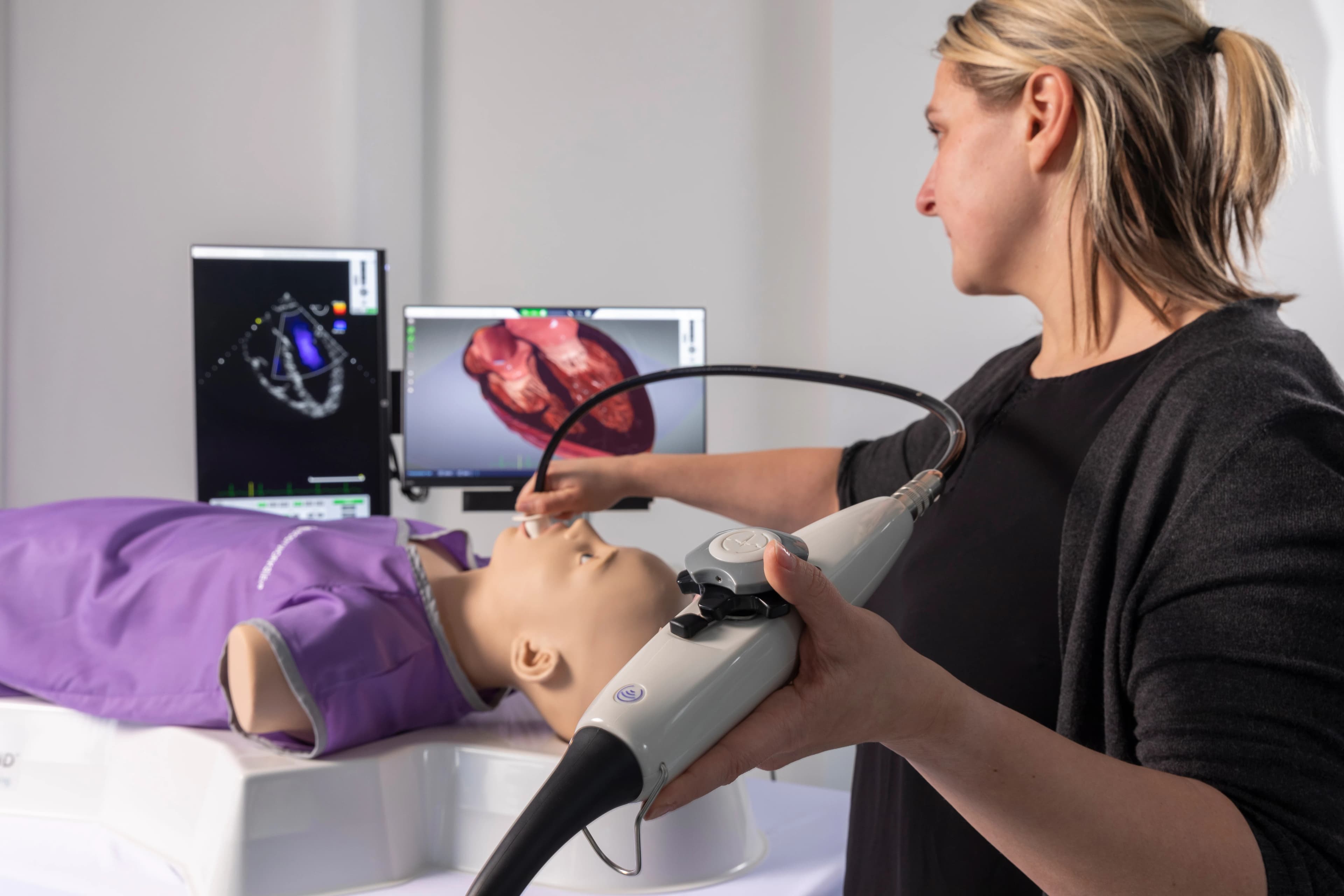
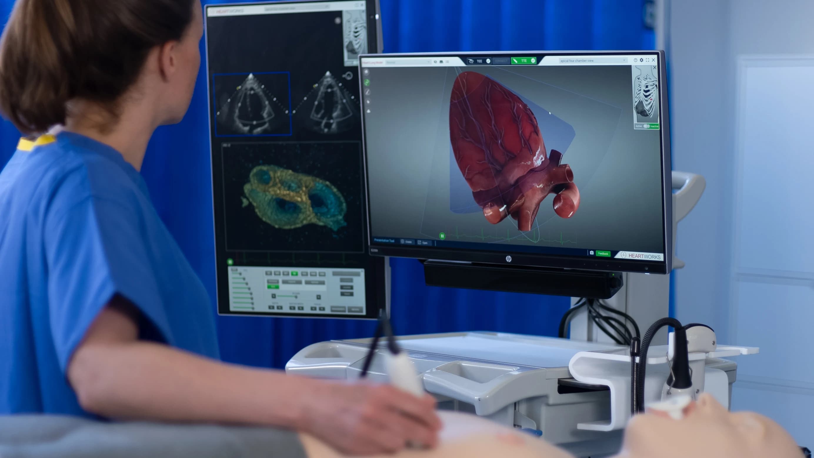
HeartWorks places the two halves side by side. On the left, the three-dimensional heart with 135 intracardiac structures, turning as the probe moves. On the right, the matching echocardiographic image. The imaging plane stops being abstract: it is the slice you watch cutting through the model.
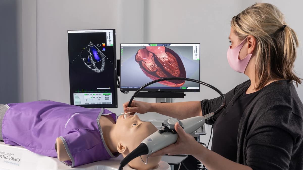
The probes are the ones used in the examination. The transthoracic rests on the manikin's chest, with anatomical landmarks for positioning; the transoesophageal carries the real controls, ante and retroflexion, lateral flexion and omniplane rotation. There are 20 transthoracic and 28 transoesophageal planes, with colour, PW and CW Doppler, and M-Mode.
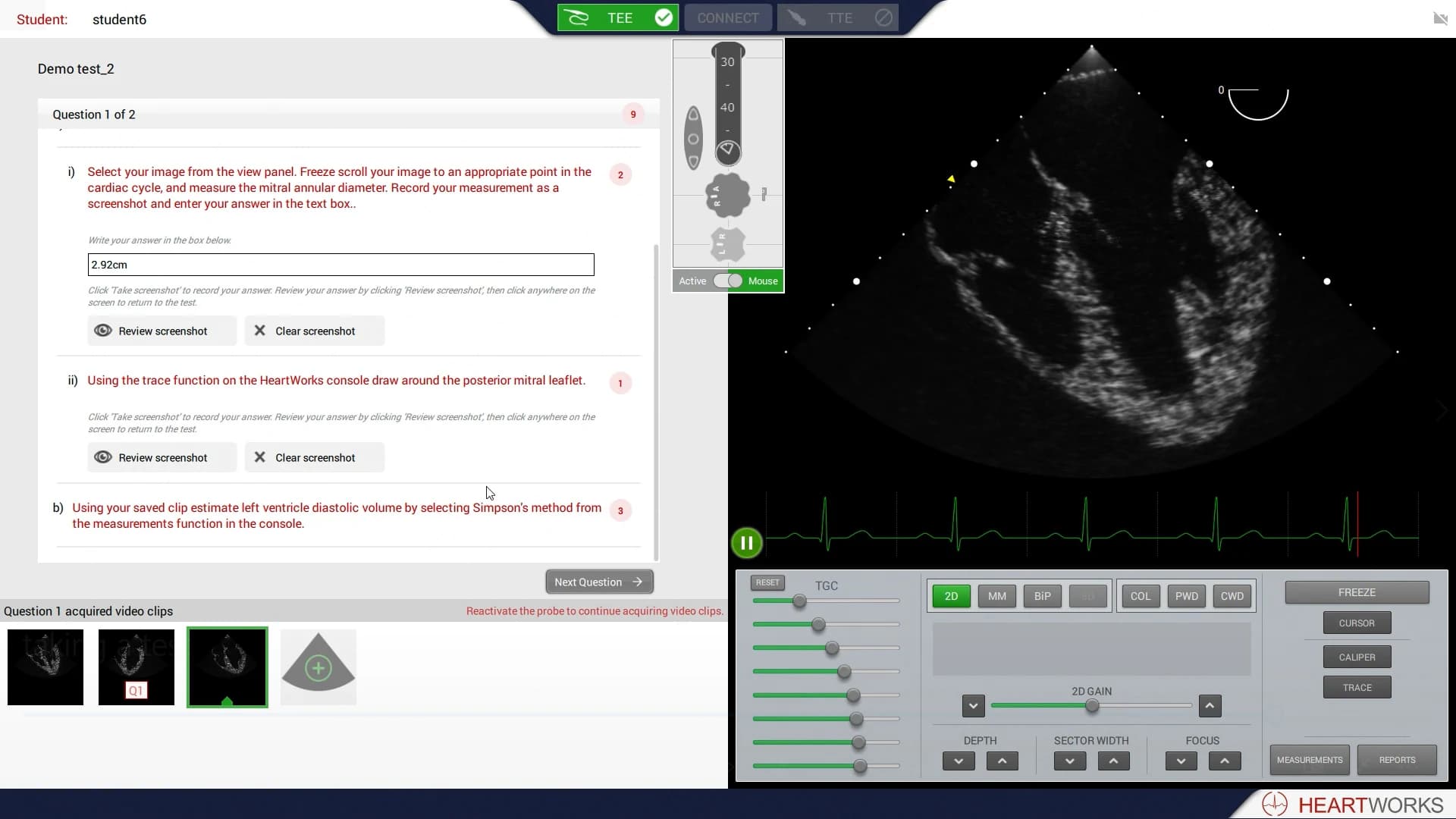
The library holds 30 patient cases, normal and diseased, including hearts with implanted devices. START 2.0 closes the loop on the teaching side: it presents the task, takes the measurement the trainee made and keeps the result for review.
What the product brings
- Interactive three-dimensional heart, with 135 intracardiac structures.
- 20 transthoracic and 28 transoesophageal imaging planes.
- Colour, PW and CW Doppler, and M-Mode.
- Real transoesophageal probe controls: ante and retroflexion, lateral flexion and omniplane rotation.
- Anatomical landmarks on the manikin for probe positioning.
- 30 patient cases, normal and diseased, including cardiac devices.
- Live 3D Echo module.
- START 2.0, to set the task, take the trainee's measurement and keep the result.
- Combined POCUS System, joining HeartWorks to BodyWorks Eve.
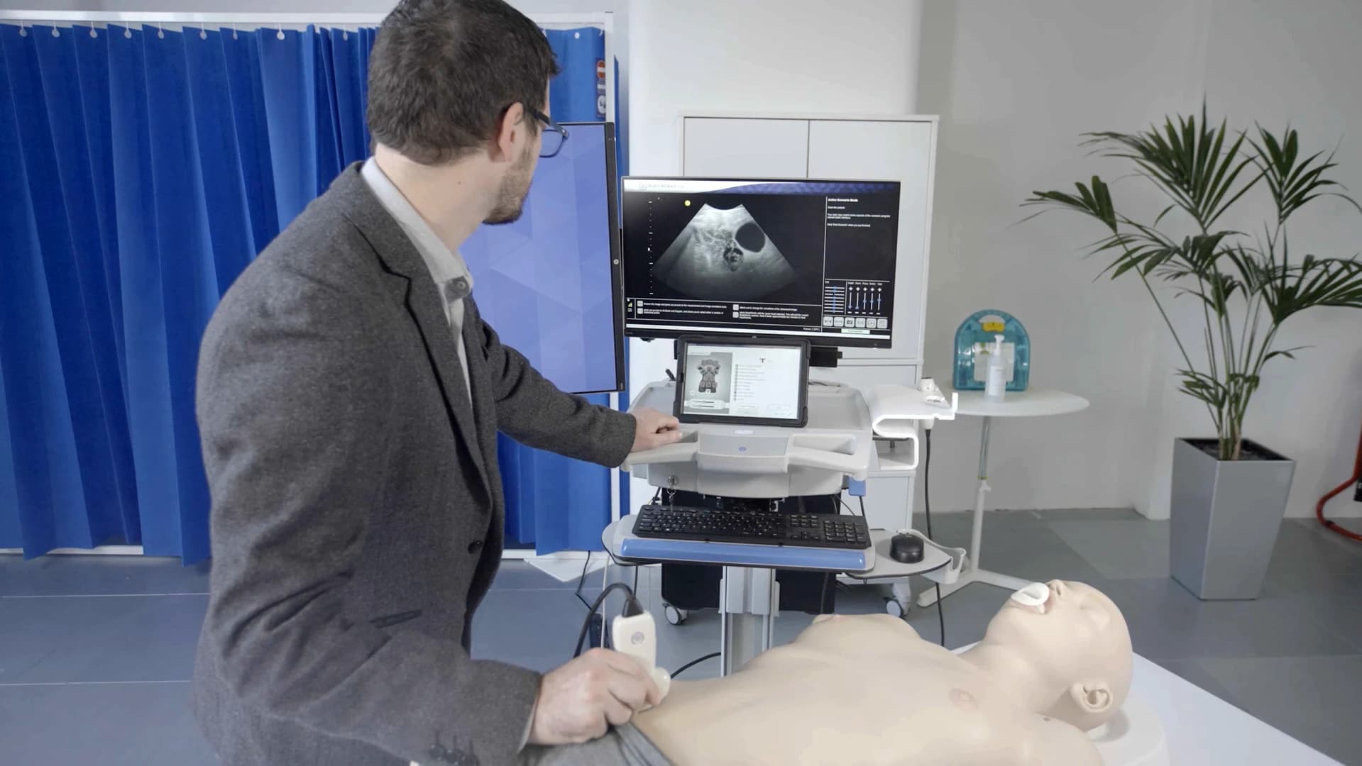
There is a tablet version too
The same three-dimensional heart exists in a tablet version, with augmented reality, that leaves the lab and goes into the hand. It is a separate product, with its own page: HeartWorks AR.
Specification
- Clinical area
- EchocardiographyCardiac anaesthesiologyCritical care
- Simulation medium
- Interactive three-dimensional heart, with 135 intracardiac structuresTransthoracic and transoesophageal probes on a manikinTEE controls: ante and retroflexion, lateral flexion and omniplane rotationAnatomical landmarks for probe positioning
- Scan modes
- 20 transthoracic imaging planes28 transoesophageal imaging planesColour, PW and CW DopplerM-Mode
- Case library
- 30 patient casesNormal and diseased states, including cardiac devices
- Modules and add-ons
- Live 3D Echo moduleSTART 2.0, student training assessment and review toolCombined POCUS System, joining HeartWorks to BodyWorks
Talk to us
Want to see HeartWorks working, or understand how it fits your department's echocardiography programme? Talk to us on WhatsApp.