

RealSpine Basic Spondy X
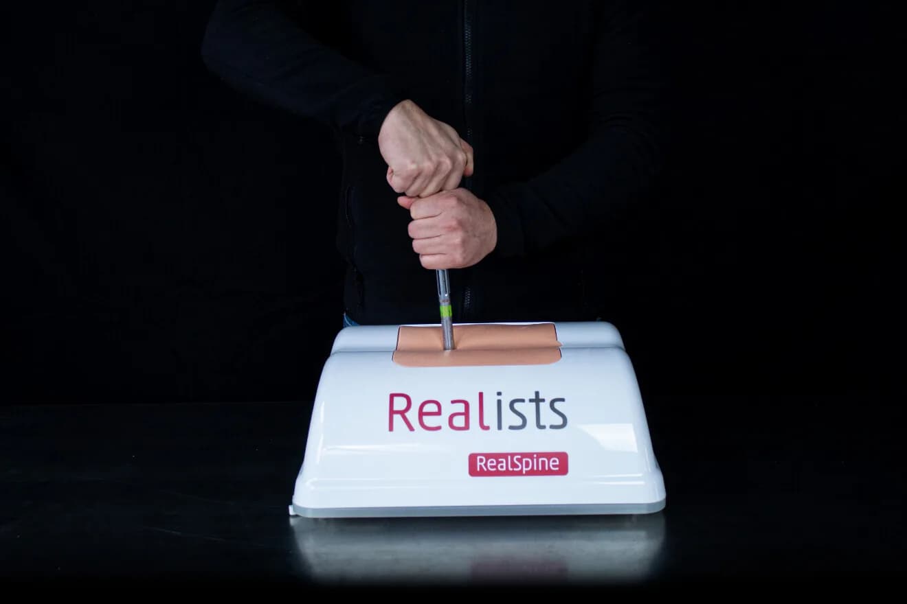
Radiolucent pedicle screw fixation simulator, L3 to S1, for training percutaneous placement with X-ray, navigation or robotics.
PresentOverview
The pedicle screw is placed almost blind. The pedicle leaves a few millimetres of margin on each side, and confirmation of the trajectory comes from the image, not from the eye.
Basic Spondy X exists for that gesture. The model is radiolucent, so the X-ray shows the instrument and the screw, not the simulator. The pathology comes ready: spondylolisthesis at L4-L5, with fixation in the pedicles of L3, L4, L5 and S1.
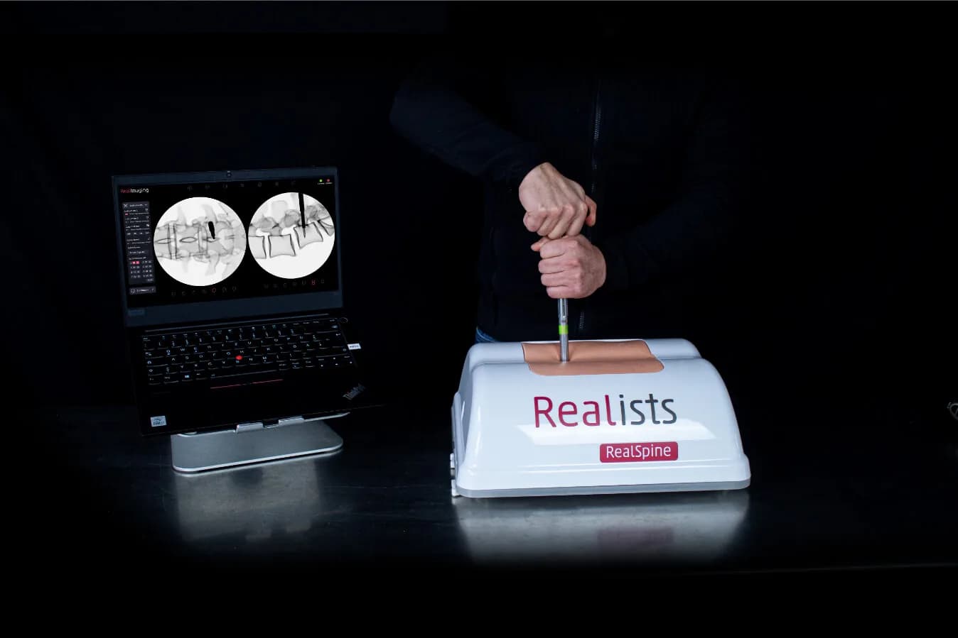
The access is posterior, open or percutaneous, and the bone and tissues keep the behaviour of the rest of the RealSpine line. The image can come from X-ray, from external navigation, from robotics, or from RealImaging when there is no C-arm in the room.
What the product brings
- Radiolucent. The image shows the instrument, not the model. That is what makes the training screen look like the surgical screen.
- Spondylolisthesis at L4-L5. The slip between the vertebrae is part of the cartridge, not of the trainee's imagination.
- Fixation at L3, L4, L5 and S1. Four pedicles to plan, the sacrum among them.
- X-ray, navigation and robotics. The same model follows all three ways of guiding the screw.
- No radiation, with RealImaging. When no C-arm is available, the image comes from navigation and nobody is exposed.
What you can train on it
- Incision
- Fusion by screw fixation at L3, L4, L5 and S1
Specification
- Approaches
- OpenPercutaneous
- Access
- Posterior
- Compatible technologies
- RealImagingExternal navigationX-rayRoboticsPedicle screws (fusion)
- Vertebral levels
- L3 to S1
- Pathologies
- Spondylolisthesis L4-L5
- Kit contents
- Reusable anatomical coverDisposable skinDisposable muscleDisposable anatomical cartridgeReusable base
Talk to us
Want to see the RealSpine Basic Spondy X up close, or clear a technical question before deciding? Talk to us right now on WhatsApp.